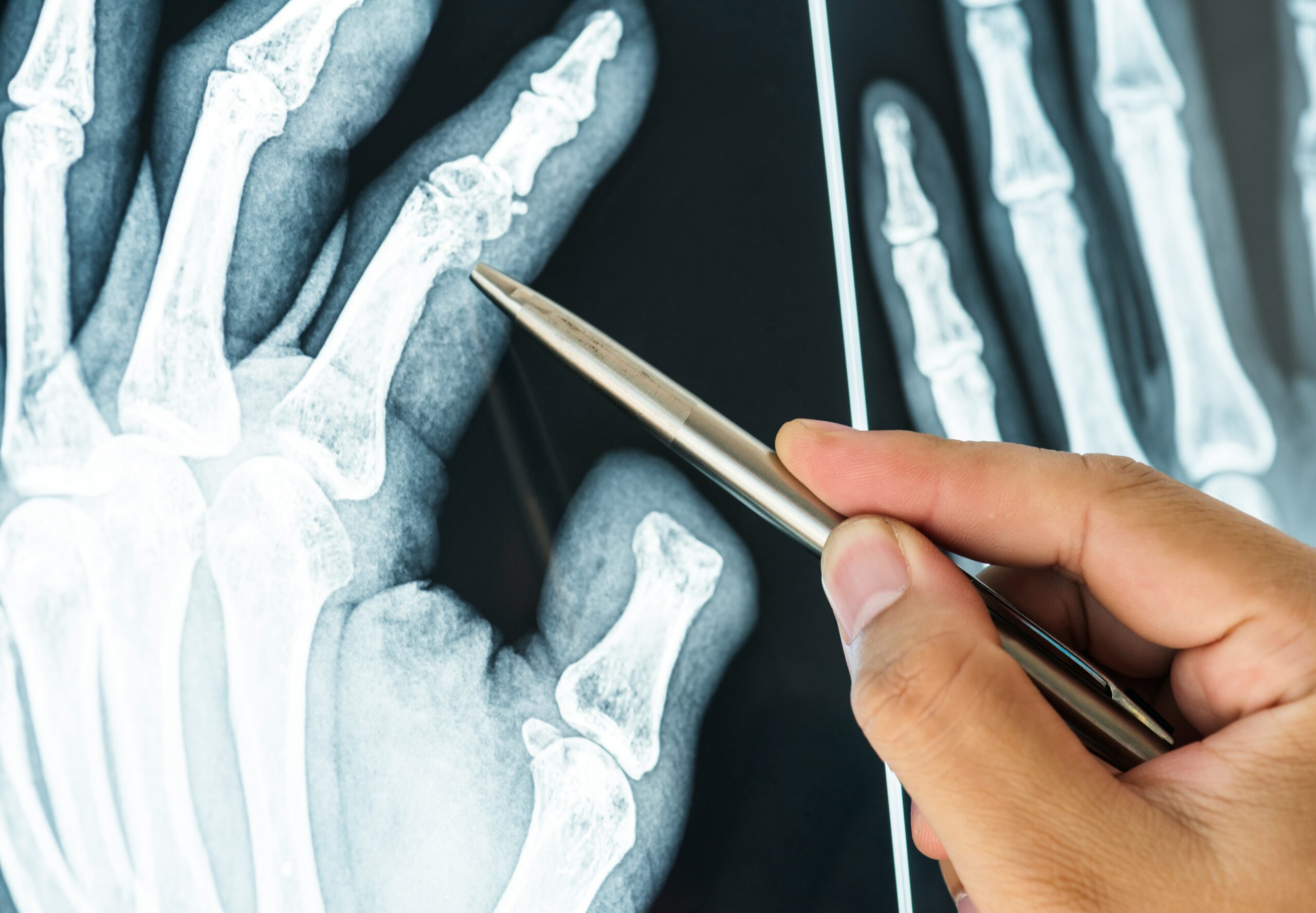
In the ever-evolving field of orthopedics, advancements in imaging technologies have played a pivotal role in enhancing diagnosis, treatment planning, and patient outcomes. As musculoskeletal disorders continue to rise due to factors such as aging populations, sports injuries, and sedentary lifestyles, the demand for precise and efficient diagnostic tools has never been greater. This article delves into the latest advancements in orthopedic imaging, highlighting innovations that are transforming patient care.
High-resolution MRI and CT Scans
Magnetic Resonance Imaging (MRI) and Computed Tomography (CT) scans have long been staples in orthopedic diagnostics, but recent advancements have significantly improved their resolution and diagnostic capabilities. High-resolution MRI technology allows for detailed imaging of soft tissues, cartilage, and ligaments, providing more precise insights into injuries and degenerative conditions. These scans now offer enhanced contrast and faster acquisition times, which not only improve the accuracy of diagnoses but also increase patient comfort by reducing scan durations.
CT scans have also evolved with the introduction of dual-energy CT (DECT) imaging. This technology uses two different energy levels to capture images, enabling better differentiation between bone and soft tissue. DECT can help identify subtle fractures and other orthopedic conditions that traditional CT may miss, allowing for more effective treatment planning.
Ultrasound Innovations
Ultrasound has gained traction in orthopedic imaging due to its accessibility, cost-effectiveness, and ability to provide real-time visualization of musculoskeletal structures. Recent advancements have led to the development of portable ultrasound devices, allowing clinicians to perform scans at the point of care. These devices are particularly beneficial for assessing soft tissue injuries, guiding injections, and monitoring rehabilitation progress.
Additionally, the integration of artificial intelligence (AI) in ultrasound imaging enhances interpretation accuracy. AI algorithms can assist in identifying abnormalities and streamline the diagnostic process, reducing the likelihood of human error and expediting patient care.
3D Imaging and Printing
Three-dimensional imaging technology has revolutionized the way orthopedic surgeons plan and execute procedures. Techniques like 3D CT and MRI allow for the creation of detailed, three-dimensional models of bones and joints. These models enable surgeons to visualize complex anatomical relationships and simulate surgical approaches before entering the operating room, leading to more precise interventions.
Moreover, advancements in 3D printing technology have facilitated the production of patient-specific implants and guides. Surgeons can now create custom implants that fit an individual’s anatomy, improving the outcomes of procedures such as joint replacements and fracture repairs. This personalization reduces the risk of complications and enhances the overall effectiveness of orthopedic surgeries.
Artificial Intelligence and Machine Learning
Artificial intelligence (AI) and machine learning are making significant inroads into orthopedic imaging, transforming the way radiologists and orthopedic surgeons interpret images. AI algorithms can analyze vast amounts of imaging data, learning to identify patterns and anomalies with remarkable accuracy. This technology not only assists in diagnosing conditions such as fractures, tumors, and degenerative diseases but also aids in predicting patient outcomes based on imaging data.
For example, AI-driven tools can evaluate MRI scans to determine the severity of cartilage damage, guiding treatment decisions for conditions like osteoarthritis. By leveraging machine learning, these systems continuously improve their diagnostic capabilities, ultimately enhancing patient care.
Enhanced Digital Imaging Systems
The integration of digital imaging systems has streamlined the workflow in orthopedic clinics and hospitals. Digital radiography allows for quicker image acquisition and immediate availability for review, enabling faster diagnosis and treatment decisions. This efficiency is crucial in emergency settings where timely interventions can significantly impact patient outcomes.
Moreover, cloud-based imaging platforms facilitate collaboration among healthcare providers. Radiologists, orthopedic surgeons, and primary care physicians can access and share imaging studies seamlessly, improving communication and ensuring that all members of a patient’s care team are on the same page. This collaborative approach enhances the overall quality of care and patient satisfaction.
Future Directions: Combining Modalities
The future of orthopedic imaging lies in the integration of multiple imaging modalities to provide a comprehensive view of musculoskeletal health. Combining MRI, CT, and ultrasound can offer a more complete assessment of orthopedic conditions, enabling more accurate diagnoses and tailored treatment plans.
Research is ongoing to develop hybrid imaging technologies that fuse the strengths of different modalities. For instance, combining functional MRI with traditional imaging can provide insights into joint mechanics and soft tissue function, allowing for a deeper understanding of conditions such as tendinopathy and ligament injuries.
The landscape of orthopedic imaging is rapidly evolving, driven by technological advancements that enhance diagnostic accuracy and improve patient outcomes. From high-resolution MRI and CT scans to innovative ultrasound devices and AI integration, these developments are reshaping the way orthopedic conditions are diagnosed and treated. As these technologies continue to advance, orthopedic surgeons will be better equipped to provide personalized and effective care, ultimately leading to improved quality of life for patients. Keeping abreast of these advancements is essential for healthcare providers to ensure they are utilizing the best tools available for their patients’ needs.